Sunday, May 7, 2017

DNA, genes and chromosomes

Your genes are part of what makes you the person you are. You are different from everyone alive now and everyone who has ever lived.

DNA

Your genes also mean that you probably look a bit like other members of your family. For example, have you been told that you have 'your mother's eyes' or 'your grandmother's nose'?

Genes influence what we look like on the outside and how we work on the inside. They contain the information our bodies need to make chemicals called proteins. Proteins form the structure of our bodies, as well playing an important role in the processes that keep us alive.

Genes are made of a chemical called DNA, which is short for 'deoxyribonucleic acid'. The DNA molecule is a double helix: that is, two long, thin strands twisted around each other like a spiral staircase.
The sides are sugar and phosphate molecules. The rungs are pairs of chemicals called 'nitrogenous bases', or 'bases' for short.

There are four types of base: adenine (A), thymine (T), guanine (G) and cytosine (C). These bases link in a very specific way: A always pairs with T, and C always pairs with G.

The DNA molecule has two important properties.
  • It can make copies of itself. If you pull the two strands apart, each can be used to make the other one (and a new DNA molecule).
  • It can carry information. The order of the bases along a strand is a code - a code for making proteins.

Genes

A gene is a length of DNA that codes for a specific protein. So, for example, one gene will code for the protein insulin, which is important role in helping your body to control the amount of sugar in your blood.

Genes are the basic unit of genetics. Human beings have 20,000 to 25,000 genes. These genes account for only about 3 per cent of our DNA. The function of the remaining 97 per cent is still not clear, although scientists think it may have something to do with controlling the genes.

Chromosomes

If you took the DNA from all the cells in your body and lined it up, end to end, it would form a strand 6000 million miles long (but very, very thin)! To store this important material, DNA molecules are tightly packed around proteins called histones to make structures called chromosomes.
Human beings have 23 pairs of chromosomes in every cell, which makes 46 chromosomes in total. A photograph of a person's chromosomes, arranged according to size, is called a karyotype.
The sex chromosomes determine whether you are a boy (XY) or a girl (XX). The other chromosomes are called autosomes.
The karyotype of a male human being
The largest chromosome, chromosome 1, contains about 8000 genes. The smallest chromosome, chromosome 21, contains about 300 genes. (Chromosome 22 should be the smallest, but the scientists made a mistake when they first numbered them!).

The DNA that contains your genes is stored in your cells in a structure called the nucleus.

A diagram of animal cell showing the nucleus
Source: University of Leicester

Saturday, April 15, 2017

Microscopic effect after stroke

We talk a lot about strokes in a clinical way in medical school. We discuss which areas of the brain are involved, and we correlate the areas damaged with the patient’s symptoms.
But what actually happens in the affected brain regions after a stroke? Injuries in the brain don’t heal like they do in other organs (you don’t form a scab and a scar in your brain). Let’s take a look at the steps the body takes to heal itself following an ischemic event in the brain.
There are basically four stages of healing following an infarct, and they usually happen in a predictable timeframe.
1. The first day (12-24 hours)
  • After brain tissue dies, it takes a while before you can see any real changes in the cells. The first changes occur in neurons. Somewhere around 12 hours following an infarct, neuronal cytoplasm develops tiny holes (microvacuoles) and takes on a deep pink-red color (the neurons are actually called red neurons at this point – you can see why in the image above). 
  • Later the nucleus undergoes pyknosis (in which it becomes small and dark) and karyorrhexis (in which it fragments into little bits, like cookie crumbs. Pathologists love food analogies and use them whenever possible.) Cells in general (but especially endothelial cells and astrocytes) tend to swell up and become more faded in color. Myelinated fibers disintegrate.

2. The second day (24-48 hours)
  • Somewhere around the end of the first day, neutrophils swarm into the area, staying until about the end of the second day, at which point they take off and are replaced by  macrophages (which come in from the blood as monocytes). Microglia (the resident phagocytic cells of the brain) become activated too. 
  • The tissue begins to undergo liquefactive necrosis from all those nasty enzymes released by the neutrophils. 
  • Macrophages are like little moms going around and cleaning up the seemingly never-ending mess. 
  • Astrocytes start to react, becoming large and getting ready to divide.

3. The next few weeks (2-3 weeks) 
  • Macrophages continue to clean stuff up. They become stuffed with debris, and you can still see some of them hanging around months or even years later. 
  • Astrocytes multiply and develop prominent, arborizing cytoplasmic extensions.

4. After several months
  • Eventually, the astrocytes calm down, and what’s left is a cavity surrounded by a dense network of glial fibers and new blood vessels. There’s no collagen formation like there is in many other organs (like skin) – so there’s no filling in of the lost tissue space.

This whole process takes place from the outside of the lesion moving inward. Which is kind of cool because you’ll often see several stages of healing going on in the same lesion.
Ref.: Pathology Student

Wednesday, February 8, 2017

Bacterial Transformation & Selection

Transfer of plasmid DNA into bacteria. How bacteria are selected. Protein production and purification.


Key points:

  • Bacteria can take up foreign DNA in a process called transformation.
  • Transformation is a key step in DNA cloning. It occurs after restriction digest and ligation and transfers newly made plasmids to bacteria.
  • After transformation, bacteria are selected on antibiotic plates. Bacteria with a plasmid are antibiotic-resistant, and each one will form a colony.
  • Colonies with the right plasmid can be grown to make large cultures of identical bacteria, which are used to produce plasmid or make protein.

The big picture: DNA cloning

Transformation and selection of bacteria are key steps in DNA cloning. DNA cloning is the process of making many copies of a specific piece of DNA, such as a gene. The copies are often made in bacteria.
In a typical cloning experiment, researchers first insert a piece of DNA, such as a gene, into a circular piece of DNA called a plasmid. This step uses restriction enzymes and DNA ligase and is called a ligation.
After a ligation, the next step is to transfer the DNA into bacteria in a process called transformation. Then, we can use antibiotic selection and DNA analysis methods to identify bacteria that contain the plasmid we’re looking for.

Steps of bacterial transformation and selection

Here is a typical procedure for transforming and selecting bacteria:


  1. Specially prepared bacteria are mixed with DNA (e.g., from a ligation).
  2. The bacteria are given a heat shock, which "encourages" them to take up a plasmid. Most bacteria do not take up a plasmid, but some do.
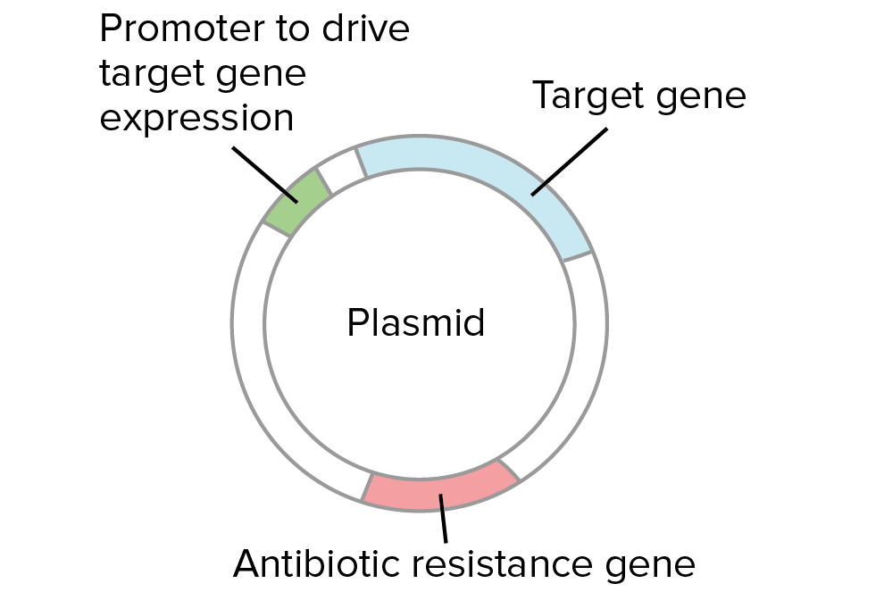
  3. Plasmids used in cloning contain an antibiotic resistance gene. Thus, all of the bacteria are placed on an antibiotic plate to select for ones that took up a plasmid.
  4. Bacteria without a plasmid die. Each bacterium with a plasmid gives rise to a cluster of identical, plasmid-containing bacteria called a colony. A typical colony looks like a small, whitish dot the size of a pinhead.
  5. Several colonies are checked to identify one with the right plasmid.
  6. A colony containing the right plasmid is grown in bulk and used for plasmid or protein production.
  1. Specially prepared bacteria are mixed with DNA (e.g., from a ligation).
  2. The bacteria are given a heat shock, which causes some of them to take up a plasmid.
    The basic answer is that a heat shock makes the bacterial membrane more permeable to DNA molecules, such as plasmids. It appears that the heat shock causes the formation of pores in the bacterial membrane, through which the DNA molecules can pass.
  3. Plasmids used in cloning contain an antibiotic resistance gene. Thus, all of the bacteria are placed on an antibiotic plate to select for ones that took up a plasmid.

Diagram of a plasmid. The plasmid contains an antibiotic resistance gene, a promoter to drive gene expression in bacteria, and the target gene inserted during the ligation.
  1. Bacteria without a plasmid die. Each bacterium with a plasmid gives rise to a cluster of identical, plasmid-containing bacteria called a colony.
  2. Several colonies are checked to identify one with the right plasmid (e.g., by PCR or restriction digest).
  1. A colony containing the right plasmid is grown in bulk and used for plasmid or protein production.

Why do we need to check colonies?

The bacteria that make colonies should all contain a plasmid (which provides antibiotic resistance). However, it’s not necessarily the case that all of the plasmid-containing colonies will have the same plasmid.
How does that work? When we cut and paste DNA, it's often possible for side products to form, in addition to the plasmid we intend to build. For instance, when we try to insert a gene into a plasmid using a particular restriction enzyme, we may get some cases where the plasmid closes back up (without taking in the gene), and other cases where the gene goes in backwards.


Left: gene goes into plasmid forwards (pointing in the same direction as the promoter sequence). This is the desired plasmid from the ligation.
Middle: plasmid closes back up without taking in the gene. This is not a useful plasmid.
Right: gene goes into plasmid backwards (pointing back towards the promoter sequence). This is not a useful plasmid if we want to express the gene in bacteria.
Let's say we are trying to insert a gene into a plasmid so it can be expressed in bacteria. In order to do so, we must "paste" the gene into the plasmid next to the promoter, pointing in the forward direction:

Starting materials:
  • Target gene digested at both ends with a particular restriction enzyme.
  • Plasmid cut with the same restriction enzyme at a site following a promoter for bacterial expression. The promoter "points" towards the right, meaning that it will drive transcription of the DNA sequence that lies to the right.
What we want to get is:
  • A recombinant plasmid where the target gene is inserted after the promoter, pointing in the forward direction (oriented so that it's transcribed to make an mRNA that specified the desired protein).
Suppose we cut our gene and plasmid with the same enzyme and join the fragments together with DNA ligase. In some cases, the plasmid DNA and the gene DNA will combine in the right way and form the plasmid we're looking for. In other cases, though, the plasmid may simply close back up (without taking in the gene), or the gene may go into the plasmid backwards. A backwards gene cannot be expressed in bacteria to make a protein.

Left: gene goes into plasmid forwards (pointing in the same direction as the promoter sequence). This is the desired plasmid from the ligation.
Middle: plasmid closes back up without taking in the gene. This is not a useful plasmid.
Right: gene goes into plasmid backwards (pointing back towards the promoter sequence). This is not a useful plasmid if we want to express the gene in bacteria.
A ligation involves many fragments of DNA (billions of copies of the plasmid, and billions of copies of the gene). Thus, in every ligation, we will get some number of "good" plasmids and some number of "bad" ones. Each colony starts from a single bacterium with a single plasmid, so all the bacteria in a colony with have the same plasmid (either "good" or "bad").
See the article on restriction enzymes and DNA ligase for a more concrete example of how and why these different ligation products can form.
Why does it matter if a gene goes into a plasmid backwards? In some cases, it doesn't. However, if we want to express the gene in bacteria to make a protein, the gene must point in the right direction relative to the promoter, or control sequence that drives gene expression. If the gene were backwards, the wrong strand of DNA would be transcribed and no protein would be made.
Because of these possibilities, it's important to collect plasmid DNA from each colony and check to see if it matches the plasmid we were trying to build. Restriction digests, PCR, and DNA sequencing are commonly used to analyze plasmid DNA from bacterial colonies.

Protein production in bacteria

Suppose that we identify a colony with a "good" plasmid. What happens next? What's the point of all that transforming, selecting, and analyzing?

Possibility 1: Bacteria = plasmid factories

In some cases, bacteria are simply used as "plasmid factories," making lots of plasmid DNA. The plasmid DNA might be used in further DNA cloning steps (e.g., to build more complex plasmids) or in various types of experiments.
In some cases, plasmids are directly used for practical purposes. For instance, plasmids were used to deliver a human gene to lung tissue in a recent gene therapy clinical trial for patients with the genetic disorder cystic fibrosisstart superscript, 1, end superscript.

Possibility 2: Bacteria = protein factories

In other cases, bacteria may be used as protein factories. If a plasmid contains the right control sequences, bacteria can be induced to express the gene it contains when a chemical signal is added. Expression of the gene leads to production of mRNA, which is translated into protein. The bacteria can then be lysed (split open) to release the protein.


A chosen colony is grown up into a large culture. The bacteria in the large culture are induced to express the target gene through addition of a chemical signal to the culture medium. Inside each bacterium, the target gene is transcribed into mRNA, and the mRNA is translated into protein. The protein encoded by the target gene accumulates inside the bacteria.
Bacteria contain many proteins and macromolecules. Because of this, the newly made protein needs to be purified (separated from the other proteins and macromolecules) before it can be used. There are a variety of different techniques used for protein purification.

Cells that have produced protein are burst open (lysed), releasing the protein and the other cell contents. The molecules extracted from the cells are applied to a column that contains antibodies specific for the target protein. Thus, the protein is trapped in the column while other molecules from the bacteria flow through. In the final step, after all the non-target proteins have been washed away, the target proteins are released from the antibodies in the column, and the pure protein is collected for use.
In one technique called affinity chromatography, a mixture of molecules extracted from the lysed bacteria is poured through a column, or a cylinder packed with beads. The beads are coated with an antibody, an immune system protein that binds specifically to a target molecule.

The antibody in the column is designed to bind to our protein of interest, and not to any other molecules in the mixture. Thus, the protein of interest is trapped in the column, while the other molecules are washed away. In the final step, the protein of interest is released from the column and collected for use.

[Source: Khan Academy]

Sunday, January 29, 2017

Neurons, Synapses, Action Potentials, and Neurotransmission

Function of neurons

The central nervous system [CNS] is composed entirely of two kinds of specialized cells: neurons and glia. Hence, every information processing system in the CNS is composed of neurons and glia; so too are the networks that compose the systems (and the maps). Clearly, without these two types of cells, the CNS would not be able to do what it does (which is everything having to do with our minds and how we move our bodies). But what do neurons and glia themselves do? What are their functions?
Neurons are the basic information processing structures in the CNS. Everything occurring above the level of neurons qualifies as information processing too. But nothing below the level of neurons does. We shall ignore that this view, called the neuron doctrine, is somewhat controversial. What isn't controversial is that the function of a neuron is to receive INPUT "information" from other neurons, to process that information, then to send "information" as OUTPUT to other neurons. (Synapses are connections between neurons through which "information" flows from one neuron to another.) Hence, neurons process all of the "information" that flows within, to, or out of the CNS. All of it! All of the motor information through which we are able to move; all of the sensory information through which we are able to see, to hear, to smell, to taste, and to touch; and of course all of the cognitive information through which we are able to reason, to think, to dream, to plan, to remember, and to do everything else that we do with our minds. Processing so many kinds of information requires many types of neurons; there may be as many as 10,000 types of them. Processing so much information requires a lot of neurons. How many? Well, "best estimates" indicate that there are around 200 billion neurons in the brain alone! And as each of these neurons is connected to between 5,000 and 200,000 other neurons, the number of ways that information flows among neurons in the brain is so large, it is greater than the number stars in the entire universe!
While we are considering numbers, it is worth noting that there are as many as 50 times more glia than neurons in our CNS! Glia (or glial cells) are the cells that provide support to the neurons. In much the same way that the foundation, framework, walls, and roof of a house prove the structure through which run various electric, cable, and telephone lines, along with various pipes for water and waste, not only do glia provide the structural framework that allows networks of neurons to remain connected, they also attend to the brain's various house keeping functions (such as removing debris after neuronal death).
Because our main interest lies in exploring how information processing occurs in the brain, we are going to ignore glia. But before we see how neurons process information (and what that means), you need to know a few things about the structure of neurons.

Structure of neurons

While there are as many as 10,000 specific types of neurons in the human brain, generally speaking, there are three kinds of neurons: motor neurons (for conveying motor information), sensory neurons (for conveying sensory information), and interneurons (which convey information between different types of neurons). The following image identifies how neurons come in various shapes and sizes. (It is based on drawings made by Cajal.)
A "typical" neuron has four distinct parts (or regions). The first part is the cell body (or soma). This is not only the metabolic "control center" of the neuron, it is also its "manufacturing and recycling plant." (For instance, it is within the cell body that neuronal proteins are synthesized.) The second and third parts are processes — structures that extend away from the cell body. Generally speaking, the function of a process is to be a conduit through which signals flow to or away from the cell body. Incoming signals from other neurons are (typically) received through its dendrites. The outgoing signal to other neurons flows along its axon. A neuron may have many thousands of dendrites, but it will have only one axon. The fourth distinct part of a neuron lies at the end of the axon, the axon terminals. These are the structures that contain neurotransmitters. Neurotransmitters are the chemical medium through which signals flow from one neuron to the next at chemical synapses.

Neuronal signaling

To support the general function of the nervous system, neurons have evolved unique capabilities for intracellular signaling (communication within the cell) and intercellular signaling (communication between cells). To achieve long distance, rapid communication, neurons have evolved special abilities for sending electrical signals (action potentials) along axons. This mechanism, called conduction, is how the cell body of a neuron communicates with its own terminals via the axon. Communication between neurons is achieved at synapses by the process of neurotransmission.

Conduction

To begin conduction, an action potential is generated near the cell body portion of the axon. An action potential is an electrical signal very much like the electrical signals in electronic devices. But whereas an electrical signal in an electronic device occurs because electrons move along a wire, an electrical signal in a neuron occurs because ions move across the neuronal membrane. Ions are electrically charged particles. The protein membrane of a neuron acts as a barrier to ions. Ions move across the membrane through ion channels that open and close due to the presence of neurotransmitter. When the concentration of ions on the inside of the neuron changes, the electrical property of the membrane itself changes. Normally, the membrane potential of a neuron rests as -70 millivolts (and the membrane is said to be polarized). The influx and outflux of ions (through ion channels during neurotransmission) will make the inside of the target neuron more positive (hence, de-polarized). When this depolarization reaches a point of no return called a threshold, a large electrical signal is generated. This is the action potential. How it is generated is illustrated in the following animation.
This signal is then propagated along the axon (and not, say, back to its dendrites) until it reaches its axon terminals. An action potential travels along the axon quickly, moving at rates up to 150 meters (or roughly 500 feet) per second. Conduction ends at the axon terminals. Axon terminals are where neurotransmission begins. Hence, it is at axon terminals where the neuron sends its OUTPUT to other neurons. At electrical synapses, the OUTPUT will be the electrical signal itself. At chemical synapses, the OUTPUT will be neurotransmitter.

Neurotransmission

Neurotransmission (or synaptic transmission) is communication between neurons as accomplished by the movement of chemicals or electrical signals across a synapse. For any interneuron, its function is to receive INPUT "information" from other neurons through synapses, to process that information, then to send "information" as OUTPUT to other neurons through synapses. Consequently, an interneuron cannot fulfill its function if it is not connected to other neurons in a network. A network of neurons (or neural network) is merely a group of neurons through which information flows from one neuron to another. The image below represents a neural network. "Information" flows between the blue neurons through electrical synapses. "Information" flows from yellow neuron A, through blue neuron B, to pink neuron C via chemical synapses.


The following animation illustrates the difference between these two kinds of synapses.

At electrical synapses, two neurons are physically connected to one another through gap junctions. Gap junctions permit changes in the electrical properties of one neuron to effect the other, and vice versa, so the two neurons essentially behave as one. Electrical neurotransmission is communication between two neurons at electrical synapses. How this occurs is explored in a bit more detail in the following animation.
Chemical neurotransmission occurs at chemical synapses. In chemical neurotransmission, the presynaptic neuron and the postsynaptic neuron are separated by a small gap — the synaptic cleft. The synaptic cleft is filled with extracellular fluid (the fluid bathing all the cells in the brain). Although very small, typically on the order of a few nanometers (a billionth of a meter), the synaptic cleft creates a physical barrier for the electrical signal carried by one neuron to be transferred to another neuron. In electrical terms, the synaptic cleft would be considered a “short” in an electrical circuit. The function of neurotransmitter is to overcome this electrical short. It does so by acting like a chemical messenger, thereby linking the action potential of one neuron with a synaptic potential in another. How this occurs is illustrated in the following animation.


Monday, November 28, 2016

microRNA

What are microRNAs?

MicroRNAs constitute a recently discovered class of non-coding RNAs that play key roles in the regulation of gene expression. Acting at the post-transcriptional level, these fascinating molecules may fine-tune the expression of as much as 30% of all mammalian protein-encoding genes.

Mature microRNAs are short, single-stranded RNA molecules approximately 22 nucleotides in length. MicroRNAs are sometimes encoded by multiple loci, some of which are organized in tandemly co-transcribed clusters.

Transcription and processing of microRNA

MicroRNA genes are transcribed by RNA polymerase II as large primary transcripts (pri-microRNA) that are processed by a protein complex containing the RNase III enzyme Drosha, to form an approximately 70 nucleotide precursor microRNA (pre-microRNA). This precursor is subsequently transported to the cytoplasm where it is processed by a second RNase III enzyme, DICER, to form a mature microRNA of approximately 22 nucleotides .The mature microRNA is then incorporated into a ribonuclear particle to form the RNA-induced silencing complex, RISC, which mediates gene silencing.
Figure: MicroRNA biogenesis

MicroRNA and gene expression

MicroRNAs usually induce gene silencing by binding to target sites found within the 3’UTR of the targeted mRNA. This interaction prevents protein production by suppressing protein synthesis and/or by initiating mRNA degradation. Since most target sites on the mRNA have only partial base complementarity with their corresponding microRNA, individual microRNAs may target as many as 100 different mRNAs. Moreover, individual mRNAs may contain multiple binding sites for different microRNAs, resulting in a complex regulatory network.

The function of microRNAs

MicroRNAs have been shown to be involved in a wide range of biological processes such as cell cycle control, apoptosis and several developmental and physiological processes including stem cell differentiation, hematopoiesis, hypoxia, cardiac and skeletal muscle development, neurogenesis, insulin secretion, cholesterol metabolism, aging, immune responses and viral replication. In addition, highly tissue-specific expression and distinct temporal expression patterns during embryogenesis suggest that microRNAs play a key role in the differentiation and maintenance of tissue identity.

MicroRNA as disease biomarkers

In addition to their important roles in healthy individuals, microRNAs have also been implicated in a number of diseases including a broad range of cancers, heart disease and neurological diseases. Consequently, microRNAs are intensely studied as candidates for diagnostic and prognostic biomarkers and predictors of drug response.

MicroRNA research

MicroRNAs were first reported in mammalian systems in 2001. In the latest release of miRBase (v.15), more than 14000 microRNAs have been annotated, highlighting the rapid growth of this field of research. However, the functions of most of these microRNAs still remain to be discovered.

The challenges of studying microRNAs are two-fold. First, microRNAs are very short (~22 nt). This means that traditional DNA-based methods are not sensitive enough to detect these sequences with any reliability. Second, closely related microRNA family members differ by as little as one nucleotide, emphasizing the need for high specificity and the ability to discriminate between single nucleotide mismatches.


Wednesday, November 16, 2016

Cancer and genes

Some genes control when new cells are made and old cells die, and repair damaged DNA. Changes (mutations) in these genes can increase the risk of cancer developing.
There are two types of mutations, called acquired mutations and inherited mutations. Acquired mutations happen during a person’s lifetime, and can’t be passed on to their children. They can happen by chance when a cell is multiplying or because a gene is damaged. Some substances, such as cigarette smoke, can increase the chance of gene mutations.
Most cancers are caused by a build-up of acquired mutations during a person’s lifetime. These are called sporadic cancers.
Inherited mutations are gene mutations that you are born with. They can be passed on to your children. Inherited gene mutations that make a cancer more likely to develop are called cancer susceptibility genes. If you inherit these genes, it doesn’t mean you’ll definitely get cancer, but you may be at an increased risk of developing it.
There are inherited cancer genes for some cancers, including breast, bowel, ovarian and womb.
Other cancers, including prostate, pancreatic and testicular, happen in some families more than usual. But specific inherited cancer genes haven’t been found for these cancers yet.

Genes and how they work

Our body is made up of tiny building blocks, called cells. Cancer develops when some cells are damaged and our body can’t repair them. The damaged cells keep growing out of the body’s control. These are cancer cells.
There are genes in every cell. All cancers are caused by changes (mutations) in genes. Genes contain the information a cell needs to work properly. This information is in a code called DNA (deoxyribonucleic acid).
Our body needs to make new cells to replace old or damaged ones. Genes control this process. They also tell cells how to repair damage. If a cell can’t be repaired, or is not needed, genes inside the cell tell it to die.
The genes that control cell growth, repair and death are called oncogenes and tumour suppressor genes. Mutations in these types of gene can increase the chance of cancer developing.

Oncogenes

These genes encourage cells to grow and multiply. A mutation in an oncogene can lead to a cell growing and multiplying out of control.

Tumour suppressor genes

These genes help protect against cancer. They control cell growth. They also repair damage to DNA.
If a cell has a mutation in a tumour suppressor gene, it may lose the ‘brakes’ on its growth. The cell can then multiply out of control.
Some tumour suppressor genes repair damage to DNA. Doctors call them DNA repair genes or caretaker genes.
When there is a mutation in a DNA repair gene, the cell can’t repair damage to itself. So cancer is more likely to develop.

Acquired mutations

Most cancers develop because of gene mutations that happen during a person’s lifetime. Doctors call these mutations acquired mutations.
Acquired gene mutations happen in the part of the body where the cancer later develops. For example, gene mutations happen in the lungs before lung cancer develops.
Many things can cause gene mutations. These include:
  • getting older
  • things in our environment such as tobacco and sunlight
  • our hormones
  • our diet.
Doctors call substances that increase the chance of gene mutations carcinogens. Radiation and the chemicals in cigarette smoke are examples of carcinogens.
Usually, several gene mutations must happen in a cell before cancer develops. This can take many years. This is why cancer is more common in older people. Cancers caused by gene mutations that happen during someone’s lifetime are called sporadic cancers.

Genes and inheritance

We inherit our genes from our parents. Everyone has two copies of each gene; one from their mother and one from their father.
Some people are born with a gene mutation that puts them at higher risk of getting cancer. Inherited mutations that make cancer more likely are called inherited cancer genes. Doctors may also call them cancer predisposition genes or cancer susceptibility genes.

If you inherit a gene mutation, it is in all your cells. This includes the sperm cells in men and the egg cells in women. So there is a 50% (1 in 2) chance of passing the gene mutation on to any children.
Inheriting a cancer gene doesn’t mean you have cancer. But, it does mean you have an increased risk of developing certain types of cancer. Further gene changes (acquired mutations) need to happen for a cancer to develop.
Doctors call cancers that develop in a family because of an inherited cancer gene inherited cancers or hereditary cancers.
Inherited cancers often develop at a younger age than sporadic cancers. Most inherited cancer genes don’t increase cancer risk until people are adults. But a few inherited cancer genes increase the risk of cancer in children. We have more information about genetic testing in children.

Can cancer genes ‘skip’ a generation?

Cancer genes cannot ‘skip’ a generation. There is a 1 in 2 (50%) chance of inheriting the gene from one of your parents. So you either inherit it or you don’t. If you don’t inherit the gene, you can’t pass it on to your children. But not everyone with the mutation develops cancer. So it can seem that the cancer skipped one generation.
The gene mutations for female cancers such as breast or ovarian cancer can pass through the father’s side of the family. Men who have the cancer gene for breast and ovarian cancer often don’t develop cancer. But they still have a 50% chance of passing the cancer gene on to their children.
If a daughter inherits a cancer gene from her father and develops breast cancer, it can seem as if the cancer gene has skipped a generation. But this isn’t the case. The mutation can’t skip a generation.

Inherited cancer genes

If a particular type of cancer occurs in a family more than in the general population, some people in the family may have an inherited cancer gene.
Scientists have found inherited cancer genes for some common cancers. These include cancers of the breast, bowel, ovary and womb.
There are other cancers that happen in some families more than usual. These include prostate, pancreatic and testicular cancers. But doctors haven’t found specific inherited cancer genes for these cancers yet.
Sometimes, there are many different types of cancer in a family. Usually, these are sporadic cancers (see above) and are due to risk factors such as age, lifestyle and the environment. But some inherited cancer genes can increase the risk of more than one type of cancer.

When cancers happen together

There are two main patterns where cancers happen together:
  • breast and ovarian cancer
  • bowel and womb cancer (sometimes with cancers of the ovary, stomach or kidney).
Other rare patterns of cancers can happen.

Low-risk genes

Not all families with more cancers than usual have an inherited cancer gene. But some families may share several genes, which increase their risk of certain cancers. These genes have a weaker effect on the risk of cancer than inherited cancer genes. They are sometimes called low-risk or low-penetrance genes.
Scientists have found several of these genes. But the effect of each gene on its own is small. And there aren’t tests available to check for them. Researchers are trying to find out how these genes interact with other risk factors to affect cancer risk.

Sunday, September 11, 2016

DNA

DNA, or deoxyribonucleic acid, is the hereditary material in humans and almost all other organisms. Nearly every cell in a person’s body has the same DNA. Most DNA is located in the cell nucleus (where it is called nuclear DNA), but a small amount of DNA can also be found in the mitochondria (where it is called mitochondrial DNA or mtDNA).
The information in DNA is stored as a code made up of four chemical bases: adenine (A), guanine (G), cytosine (C), and thymine (T). Human DNA consists of about 3 billion bases, and more than 99 percent of those bases are the same in all people. The order, or sequence, of these bases determines the information available for building and maintaining an organism, similar to the way in which letters of the alphabet appear in a certain order to form words and sentences.
DNA bases pair up with each other, A with T and C with G, to form units called base pairs. Each base is also attached to a sugar molecule and a phosphate molecule. Together, a base, sugar, and phosphate are called a nucleotide. Nucleotides are arranged in two long strands that form a spiral called a double helix. The structure of the double helix is somewhat like a ladder, with the base pairs forming the ladder’s rungs and the sugar and phosphate molecules forming the vertical sidepieces of the ladder.
An important property of DNA is that it can replicate, or make copies of itself. Each strand of DNA in the double helix can serve as a pattern for duplicating the sequence of bases. This is critical when cells divide because each new cell needs to have an exact copy of the DNA present in the old cell.
DNA is a double helix formed by base pairs attached to a sugar-phosphate backbone.
Credit: U.S. National Library of Medicine

Gene

A gene is the basic physical and functional unit of heredity. Genes, which are made up of DNA, act as instructions to make molecules called proteins. In humans, genes vary in size from a few hundred DNA bases to more than 2 million bases. The Human Genome Project has estimated that humans have between 20,000 and 25,000 genes.
Every person has two copies of each gene, one inherited from each parent. Most genes are the same in all people, but a small number of genes (less than 1 percent of the total) are slightly different between people. Alleles are forms of the same gene with small differences in their sequence of DNA bases. These small differences contribute to each person’s unique physical features.

Genes are made up of DNA. Each chromosome contains many genes.
Credit: U.S. National Library of Medicine


Saturday, September 12, 2015

GENE: The Ultimate Fact

What are genes?

Genes are pieces of DNA (deoxyribonucleic acid) inside each cell that tell the cell what to do and when to grow and divide. Each gene is made up of a specific DNA sequence that contains the code (the instructions) to make a certain protein, each of which has a specific job or function in the body. Each human cell has about 25,000 genes.
Most genes are contained in chromosomes. A chromosome is a long strand of DNA wrapped around a special protein called histone. Most chromosomes contain many different genes. Most human cells contain 23 pairs of chromosomes – one pair of sex chromosomes (either XX in females or XY in males) plus 22 pairs of non-sex chromosomes called autosomes. Sperm and egg cells only contain half as many chromosomes (23). Chromosomes are passed from parents to their children through sperm and egg cells. One chromosome of each pair is inherited from the mother, and the other comes from the father. This is why children look like their parents, and why they may have a tendency to develop certain diseases that run in their families.
A cell uses its genes selectively; that is, it can turn on (or activate) the genes it needs at the right moment and turn off other genes that it doesn't need. All the cells in the body (except egg and sperm) contain the same genes. Turning on some genes and turning off others is how a cell becomes specialized. That is how a cell becomes a muscle cell and not a bone cell, for example. Some genes stay active all the time to make proteins needed for basic cell functions. Others shut down when their job is finished and start again later if needed.

Dominant vs. recessive genes

We have 2 versions (copies) of most genes – one from each parent. For some versions of a gene, only one copy is needed to see a certain quality or disease (in genetics this is called a trait). These genes are called dominant. If both copies have to be the same to see that trait, it is called recessive. For example, the gene for brown eyes is dominant while the gene for blue eyes is recessive, so if you get one copy of the brown eye gene from one parent and a copy of the blue eye gene from the other, you will have brown eyes. You will only get blue eyes if you get 2 copies of the blue eye gene (one from each parent). This classification applies to gene mutations as well. If you only need to inherit one copy of a gene mutation to get a disease or syndrome, it is called dominant. If you need 2, it is called recessive. 
X-linked genes
Things are a little different in terms of genes on the X chromosome. Normally, we each have 2 sex chromosomes. Women have two X chromosomes, while males have one X chromosome and one Y chromosome. Since the Y chromosome contains different genes than the X chromosome, males have only one copy of the genes on the X chromosome. Some diseases/conditions are caused by genes on the X chromosome. For some of these, like color blindness, a female has to have 2 copies of the gene (one on each X chromosome) to get the condition. For a male though, he only has to have the gene on his one X chromosome. Diseases and conditions like this are called X-linked. X-linked conditions are more common in males.

Gene mutations

Mutations are abnormal changes in the DNA of a gene. The building blocks of DNA are called bases. The sequence of the bases determines the gene and its function. Mutations involve changes in the arrangement of the bases that make up a gene. Even a change in just one base among the thousands of bases that make up a gene can have a major effect.
A gene mutation can affect the cell in many ways. Some mutations stop a protein from being made at all. Others may change the protein that is made so that it no longer works the way it should or it may not even work at all. Some mutations may cause a gene to be turned on, and make more of the protein than usual. Some mutations don't have a noticeable effect, but others may lead to a disease. For example, a certain mutation in the gene for hemoglobin causes the disease sickle cell anemia.
Cells become cancer cells largely because of mutations in their genes. Often many mutations are needed before a cell becomes a cancer cell. The mutations may affect different genes that control cell growth and division. Some of these genes are called tumor suppressor genes. Mutations may also cause some normal genes to become cancer-causing genes known as oncogenes.
We have 2 copies of most genes, one from each chromosome in a pair. In order for a gene to stop working completely and potentially lead to cancer, both copies have to be “knocked out” with mutations. That means for most genes, it takes 2 mutations to make that gene stop working completely.

Types of mutations

There are 2 major types of gene mutations, inherited and acquired:
  • An inherited gene mutation is present in the egg or sperm that formed the child. After the egg is fertilized by the sperm, it created one cell called a zygote that then divided to create a fetus (which became a baby). Since all the cells in the body came from this first cell, this kind of mutation is in every cell in the body (including some eggs or sperm) and so can be passed on to the next generation. This type of mutation is also called germline (because the cells that develop into eggs and sperm are called germ cells) or hereditary. Inherited mutations are thought to be a direct cause of only a small fraction of cancers.
  • An acquired mutation is not present in the zygote, but is acquired some time later in life. It occurs in one cell, and then is passed on to any new cells that are the offspring of that cell. This kind of mutation is not present in the egg or sperm that formed the fetus, so it cannot be passed on to the next generation. Acquired mutations are much more common than inherited mutations. Most cancers are caused by acquired mutations. This type of mutation is also called sporadic, or somatic.

Mutations and cancer

Experts agree that it takes more than one mutation in a cell for cancer to occur. When someone has inherited an abnormal copy of a gene, though, their cells already start out with one mutation. This makes it all the easier (and quicker) for enough mutations to build up for a cell to become cancer. That is why cancers that are inherited tend to occur earlier in life than cancers of the same type that are not inherited.
Even if you were born with healthy genes, some of them can become changed (mutated) over the course of your life. These acquired mutations cause most cases of cancer. Some acquired mutations can be caused by things that we are exposed to in our environment, including cigarette, smoke, radiation, hormones, and diet. Other mutations have no clear cause, and seem to occur randomly as the cells divide. In order for a cell to divide to make 2 new cells, it has to copy all of its DNA. With so much DNA, sometimes mistakes are made in the new copy (like typos). This leads to DNA changes (mutations). Every time a cell divides, it is another opportunity for mutations to occur. The numbers of gene mutations build up over time, which is why we have a higher risk of cancer as we get older.
It is important to realize that gene mutations happen in our cells all the time. Usually, the cell detects the change and repairs it. If it can’t be repaired, the cell will get a signal telling it to die in a process called apoptosis. But if the cell doesn't die and the mutation is not repaired, it may lead to a person developing cancer. This is more likely if the mutation affects a gene involved with cell division or a gene that normally causes a defective cell to die.
Some people have a high risk of developing cancer because they have inherited mutations in certain genes.

Penetrance

For dominant genes and mutations, the term penetrance is used to indicate the proportion of those carrying a mutation who will have the trait, syndrome, or disease. If all of the people who inherit the mutation have the disease, it is called complete penetrance. If not all of the people who have the mutation get the disease, it is called incomplete penetrance. In general, inherited mutations leading to cancer have incomplete penetrance, meaning not everyone with the mutation will get cancer. That is in part because although the person has a mutation in one copy of the gene, he or she needs to acquire at least one more mutation for the gene to stop working completely and cancer to occur. Since not everyone gets the second mutation, not everyone gets cancer. Incomplete penetrance can also be because even if the mutation makes it so that a gene doesn’t function, other factors may be needed for the cancer to start.
High vs. low penetrance
Gene mutations can cause large changes in the function of a gene. They may even cause that copy of the gene to stop working altogether. When an inherited mutation has a large enough effect on the function of a gene to cause a disease or noticeable problem in most of the people who have it, that mutation is called “high penetrance.”
High-penetrance mutations in cancer susceptibility genes can lead to many people in a family getting certain kinds of cancers – a family cancer syndrome. These are thought to cause only a small fraction of cancers that run in a family. For example, only about 1/5 of the breast cancer that runs in families is thought to be caused by high-penetrance mutations in genes like BRCA1 and BRCA2.
Some inherited mutations, though, don’t seem to affect gene function very much and don’t often cause obvious problems. These mutations are called “low-penetrance.” Low-penetrance mutations can affect cancer risk through subtle effects on things like hormone levels, metabolism, or other things that interact with risk factors for cancer. Low-penetrance mutations, together with gene variants (discussed below) are thought to be responsible for most of the cancer risk that runs in families.

Gene variants

People can also have different versions of genes that are not mutations. Common differences in genes are called variants. These versions are inherited and are present in every cell of the body. The most common type of gene variant involves a change in only one base (nucleotide) of a gene. These are called single nucleotide polymorphisms (SNPs, pronounced “snips”). There are estimated to be millions of SNPs in each person’s DNA.
Other types of variants are less common. Many genes contain sequences of bases that are repeated over and over. A common type of variant involves a change in the number of these repeats.
Some variants have no apparent effect on the function of the gene. Others tend to influence the function of genes in a subtle way, such as making them slightly more or less active. These changes don’t cause cancer directly, but can make someone more likely to get cancer by affecting things like hormone levels and metabolism. For example, some gene variants affect levels of estrogen and progesterone, which can affect the risk of breast and endometrial cancers. Others can affect the breakdown of toxins in cigarette smoke, making a person more likely to get lung and other cancers.
Gene variants can also play a role in diseases that impact cancer risk – like diabetes and obesity.
Variants and low-penetrance mutations can be similar. The main difference between the two is how common they are. Mutations are rare, while gene variants are more common.
Still, since these variants are common and someone can have many of them, their effect can add up. Studies have shown that these variants can influence cancer risk and, together with low penetrance mutations, they may account for a large part of the cancer risk that runs in families.

Other ways cells change genes and gene activity

Although all of the cells of your body contain the same genes (and DNA), different genes are active in some cells than in others. Even within a certain cell, some genes are active at some times and inactive at others. Turning on and off of genes in this case isn’t based on changes in the DNA sequence (like mutations), but by other means calledepigenetic changes.
DNA methylation: In this type of epigenetic change, a molecule called a methyl group is attached to certain nucleotides. This changes the structure of the DNA so that the gene can’t start the process of making the protein for which it codes (this process is called transcription). This basically turns off the gene. In some people with a mutation in one copy of a cancer susceptibility gene, the other copy of the gene becomes inactive not by mutation, but by methylation.
Histone modification: Chromosomes are made up of DNA wrapped around proteins called histones. Histone proteins can be changed by adding (or subtracting) something called an acetyl group. Adding acetyl groups (acetylation) can activate (turn on) that part of the chromosome, while taking them away (deacetylation) can deactivate it (turn it off). Methylation is also used to activate and deactivate parts of chromosomes. Histone proteins can also be changed by adding or subtracting methyl groups (methylation and demethylation). Although abnormal histone modification isn’t known to cause cancer, drugs that alter histone modifications can help in the treatment of cancer by turning on genes that help control cell growth and division.
RNA interference: RNA (ribonucleic acid) is important inside cells as the middle step that allows genes to code for proteins. But some small forms of RNA can interfere with gene expression by attaching to other pieces of RNA, or even affecting histones or DNA itself. Drugs are being developed that affect abnormal genes in cancer cells through RNA interference.

Two of the main types of genes that play a role in cancer are oncogenes and tumor suppressor genes.

Oncogenes

Proto-oncogenes are genes that normally help cells grow. When a proto-oncogene mutates (changes) or there are too many copies of it, it becomes a "bad" gene that can become permanently turned on or activated when it is not supposed to be. When this happens, the cell grows out of control, which can lead to cancer. This bad gene is called an oncogene.
It may be helpful to think of a cell as a car. For it to work properly, there need to be ways to control how fast it goes. A proto-oncogene normally functions in a way that is much like a gas pedal. It helps the cell grow and divide. An oncogene could be compared with a gas pedal that is stuck down, which causes the cell to divide out of control.
A few cancer syndromes are caused by inherited mutations of proto-oncogenes that cause the oncogene to be turned on (activated). But most cancer-causing mutations involving oncogenes are acquired, not inherited. They generally activate oncogenes by:
  • Chromosome rearrangements: Changes in chromosomes that put one gene next to another, which allows one gene to activate the other
  • Gene duplication: Having extra copies of a gene, which can lead to it making too much of a certain protein

Tumor suppressor genes

Tumor suppressor genes are normal genes that slow down cell division, repair DNA mistakes, or tell cells when to die (a process known as apoptosis or programmed cell death). When tumor suppressor genes don't work properly, cells can grow out of control, which can lead to cancer.
A tumor suppressor gene is like the brake pedal on a car. It normally keeps the cell from dividing too quickly, just as a brake keeps a car from going too fast. When something goes wrong with the gene, such as a mutation, cell division can get out of control.
An important difference between oncogenes and tumor suppressor genes is that oncogenes result from the activation (turning on) of proto-oncogenes, but tumor suppressor genes cause cancer when they are inactivated(turned off).
Inherited abnormalities of tumor suppressor genes have been found in some family cancer syndromes. They cause certain types of cancer to run in families. But most tumor suppressor gene mutations are acquired, not inherited.
For example, abnormalities of the TP53 gene (which codes for the p53 protein) have been found in more than half of human cancers. Acquired mutations of this gene appear in a wide range of cancers.

Cancer diagnosis and monitoring treatment

Certain mutations are commonly found in the cells of some types of cancers. Finding certain mutations in cells can confirm the diagnosis of that cancer. Testing cells for the mutation can also be used after diagnosis to see how the cancer is responding to treatment.
For example, the leukemia cells of patients with chronic myeloid leukemia (CML) contain a mutated gene called BCR-ABL. In order to be diagnosed with CML, this mutation must be present, so testing for this mutation is used to confirm the diagnosis. Very sensitive tests can tell how many copies of this mutation are present in a blood sample (which indicates how many CML cells are present). These tests can find even tiny amounts, representing small numbers of CML cells among millions of normal cells. The number of copies is determined when treatment is started, and then again sometime later to see how well the treatment is working. If treatment has put the leukemia into remission, this test can be used to see if it is coming back and new treatment is needed.

Genes and cancer prognosis

In some cancers, specific gene changes can be used to predict which patients are likely to have a better or worse outcome. This can help guide the intensity of treatment.
For example, patients with acute myeloid leukemia (AML) whose leukemia cells have a mutation in the FLT3 gene have a poorer prognosis than patients whose leukemia cells do not contain that mutation. Doctors may recommend more intense treatment, includingstem cell transplant for someone whose leukemia cells have this mutation. On the other hand, people whose leukemia cells have mutations in the NPM1 gene (and no other abnormalities) seem to have a better prognosis than people without this mutation. As a result, doctors may not feel that a stem cell transplant is needed in someone whose leukemia cells only have a NPM1 mutation.
For some cancers, tests that look at the activity (expression) of many genes at once can be useful in predicting prognosis. These tests, called gene expression panels, are performed on samples of the cancer. They are available for a number of cancers, including breast, colon, and prostate cancers. These tests can help predict which patients are more likely to have their cancers come back after treatment. So far, though, only one, the Oncotype Dx® breast cancer assay, has been shown to help predict which patients benefit the most from certain treatments.

Genes and cancer treatment

Drugs targeting genes or gene mutations

Drugs have been developed that target some of the gene changes in certain cancers. Actually these drugs often target the protein made by the abnormal gene (and not the gene itself).
For example, HER2/neu is a proto-oncogene in normal cells that helps them grow. It becomes an oncogene when a cell has too many copies of this gene. When this happens, the cells make too much HER2/neu protein and the cancer is said to be HER2 positive. Patients with breast cancer with cells that are HER2 positive do not respond as well to certain chemotherapy drugs. But newer drugs such as trastuzumab, lapatinib, and several others, have been designed to specifically attack cells that are HER2 positive. These drugs can slow cancer cell growth and improve outcomes in patients with HER2 positive cancers. Breast cancers are now routinely tested to see if they are or the HER2 positive to identify which patients will benefit from these drugs. Other cancers also can be HER2 positive. Anti-HER2 therapy has also helped people with stomach cancer that is HER2-positive.
In chronic myeloid leukemia (CML), the cancer cells have a gene change called BCR-ABL that makes a type of protein called a tyrosine kinase. Drugs that target the BCR-ABL protein, such as imatinib, are often very effective against CML. They lead to remission of the leukemia in most patients treated in the early stages of their disease.
Drugs targeting certain mutations are useful in a number of other cancers including acute lymphocytic leukemia, gastrointestinal stromal tumors, non-small cell lung cancer, a certain kind of non-Hodgkin lymphoma, and melanoma.

Drugs that activate genes

DNA methylation is one way to turn-off genes. Drugs called hypomethylating agents can reverse methylation. This can be helpful in treating some cancers in which some genes are abnormally methylated. For example, in myelodyplastic syndrome, certain genes that are often methylated in the cancer cells when they aren’t supposed to be. The hypomethylating agents decitabine and azacytidine can decrease this abnormal methylation, which can be useful in treating this disease.
Other drugs that help fight cancer by activating genes are the histone deacetylase inhibitors, such as vorinostat and romidepsin .

Gene testing to help predict if a drug will work

Some drugs don’t help patients if the cancer cells have certain gene mutations. For example, cetuximab and panitumumab are drugs used to treat advanced colorectal cancers. However, these drugs don’t help patients with cancers that have mutations in the KRAS gene, so doctors check the cancer cells for these mutations before they give either of these drugs.
Some drugs work better in people with certain mutations. For example, the drug erlotinib , which can be used to treat non-small cell lung cancer, works better in patients whose cancer cells have a certain mutation in the EGFR gene.

Future directions

Many researchers are very hopeful about the future of cancer treatments based on the specific gene changes found in cancer cells, and this remains a very active area of research. There are many clinical trials under way today that could lead to better treatments for many types of cancer.