Monday, September 23, 2019

Macrophage – the new miracle cell? How the phagocytic cell helps maintaining a healthy heart beat

For some people, when they think of a macrophage, they will imagine a real life microscopic version of Pac-Man running around their body ingesting any microbial intruder lying in its way. While this has been the central dogma surrounding this immune cell for the last 100 years - it is becoming increasingly more obvious that this isn’t the entire truth. Within the last 10 years, numerous papers have been published identifying new functions and processes carried out by the humble macrophage. More importantly, we have shown that tissue resident macrophages carry out organ-specific functions. For instance, macrophages have been shown to contribute to iron recycling in the spleen and liver (Theurl et al. 2016), thermogenesis regulation in adipose tissue (Nguyen et al. 2011) and more recently, electrical conduction in the heart (Hulsmans et al. 2017). 
Are you trying to tell me that these tiny white blood cells are better at multitasking than I am? Well….Yes. That is exactly what I am telling you.
study published in Cell by a team of researchers from Harvard Medical School, revealed that cardiac-resident macrophages play an important role in facilitating electrical conduction in the steady state heart.
By coupling cardiac research techniques such as surface and telemetric ECG monitoring with immunological techniques like flow cytometry and the use of transgenic mice, they uncovered the secret life of a cardiac macrophage.

Cardiac macrophages form Cx43-containing gap junctions with neighbouring cardiomyocytes


Cardiomyocytes, or heart cells, communicate with one another via gap junctions. Gap junctions allow for the transmission of small molecules and ions between neighbouring cells, which in the case of the heart, facilitates the synchronous contraction of the tissue and subsequent generation of a heartbeat. Connexin (Cx) proteins are the nuts and bolts of these intercellular channels and can come in various isoforms. The particular isoform Cx43, has been shown to connect cardiomyocytes with non-cardiomyocytes.
Using Flow cytometry and RNA sequencing, the team were able to show that AV node resident macrophages express Cx43. To prove that the macrophages were using the Cx43 connexin proteins to form gap junctions with neighbouring cardiac cells, they took a closer look at the cells using electron microscopy. This allowed the team to visualize direct membrane-membrane contact between the two cell types, indicating possible cell-cell communication.

Macrophages influence AV node conduction

To investigate whether cell-cell communication was happening through the Cx43-junctions, the team used a loss-of-function experiment where they deleted Cx43 in AV node resident macrophages. To do this, they used a tamoxifen inducible system, where by injecting the transgenic mice with tamoxifen, they could specifically delete Cx43 in the cardiac macrophages. To measure the effects of removing the Cx43 junctions on AV node conduction, they performed an in vivo electrophysiology (EP) study using a Millar Mikro-Tip octapolar catheter (EPR-800) inserted into the right atrium and ventricle in conjunction with surface ECG/EKG recordings.
Surface ECG recordings we measured using subcutaneous electrodes connected to an Animal Bio amplifier and PowerLab. Data was later analyzed using the ECG module in LabChart Pro. This enabled the team to stimulate the heart in different ways and measure the subsequent effects on the electrical activity of the heart. Comparing results from the control and transgenic mice, they found that in the absence of Cx43, there was impaired AV node conduction, indicating that cardiac macrophages can influence the electrical conduction of the heart.


ECG telemetry reveals mice lacking macrophages have irregular heart beats 

Next the team wanted to investigate if depleting the resident macrophage population would have a similar effect on AV-node conduction. They did this by genetically modifying mice to expressess a diphtheria toxin (DT) inducible system, that upon administration of DT, would result in the depletion of macrophages (and other myeloid cells). To monitor the subsequent effects on the heart, they used an implantable ECG telemetry device (DSI) placed in a lead II position within the abdomen. This enabled ECG data to be recorded continuously over 8 days. The data was later analyzed using LabChart software. Interestingly, within 24 hours of a single dose of DT, all mice had developed a first degree AV block, that overtime progressed to a third degree block. An AV-block happens when the conduction between the atria and ventricles is impaired. Clinically, AV-blocks can worsen pre-existing conditions such as heart failure.
The unique integration of cardiac and immunological research techniques used in this study, has uncovered the unlikely role that macrophages play in facilitating electrical conduction in the heart and their potential involvement in heart disease. For years we have compartmentalized our bodies into distinct physiological systems, made up of different cell types that have distinct functions. However, it is through such studies like this that we are beginning to understand the amazing adaptability and cooperativity of these so called ‘distinct’ cell types, and discover how intertwined these systems really are.

Courtesy: Adinstruments

Wednesday, April 10, 2019

Types of Monocytes

Monocytes are a group of immune cells that originate in bone marrow and are released into peripheral blood, where they circulate for several days. They belong to the mononuclear-phagocyte system, which also include macrophages, dendritic cells, and their bone-marrow precursors. Monocytes represent 5–10% of peripheral leucocytes .

Monocytes have been divided into three subtypes based on relative surface expression of LPS co-receptor CD14 and FCγIII receptor CD16. The most predominant of the three, termed “classical monocytes”, express high levels of CD14 on their surface, are devoid of surface CD16, and account for approximately 80% of the total monocyte population. The remaining 20% express CD16 and have been further classified into two subtypes. The more abundant “nonclassical monocytes”, are characterized by very low expression of surface CD14 and high levels of CD16, whereas the third monocyte subtype, called “intermediate monocytes”, express high levels of both the receptors.

Wednesday, February 6, 2019

Protein targeting

Different proteins need to be sent to different parts of a eukaryotic cell, or, in some cases, exported out of the cell and into the extracellular space. How do the right proteins get to the right places?
Cells have various shipping systems, kind of like molecular versions of the postal service, to make sure that proteins arrive at their correct destinations. In these systems, molecular labels (often, amino acid sequences) are used to "address" proteins for delivery to specific locations. Let’s take a look at how these shipping systems work.

Overview of cellular shipping routes

Translation of all proteins in a eukaryotic cell begins in the cytosol (except for a few proteins made in mitochondria and chloroplasts). As a protein is made, it passes step by step through a shipping "decision tree." At each stage, the protein is checked for molecular tags to see if it needs to be re-routed to a different pathway or destination.
Diagram based on similar diagram in Alberts et al. ^1
The first major branch point comes shortly after translation starts. At this point, the protein will either remain in the cytosol for the rest of translation, or be fed into the endoplasmic reticulum (ER) as it is translated^2.
  • Proteins are fed into the ER during translation if they have an amino sequence called a signal peptide. In general, proteins bound for organelles in the endomembrane system (such as the ER, Golgi apparatus, and lysosome) or for the exterior of the cell must enter the ER at this stage.
  • Proteins that do not have a signal peptide stay in the cytosol for the rest of translation. If they lack other "address labels," they'll stay in the cytosol permanently. However, if they have the right labels, they can be sent to the mitochondria, chloroplasts, peroxisomes, or nucleus after translation. 

The endomembrane system and secretory pathway

Proteins destined for any part of the endomembrane system (or the outside of the cell) are brought to the ER during translation and fed in as they're made.

Signal peptides

The signal peptide that sends a protein into the endoplasmic reticulum during translation is a series of hydrophobic (“water-fearing”) amino acids, usually found near the beginning (N-terminus) of the protein. When this sequence sticks out of the ribosome, it’s recognized by a protein complex called the signal-recognition particle (SRP), which takes the ribosome to the ER. There, the ribosome feeds its amino acid chain into the ER lumen (interior) as it's made.
In some cases, the signal peptide is snipped off during translation and the finished protein is released into the interior of the ER (as shown above). In other cases, the signal peptide or another stretch of hydrophobic amino acids gets embedded in the ER membrane. This creates a transmembrane (membrane-crossing) segment that anchors the protein to the membrane.

Transport through the endomembrane system

In the ER, proteins fold into their correct shapes, and may also get sugar groups attached to them. Most proteins are then transported to the Golgi apparatus in membrane vesicles. Some proteins, however, need to stay in the ER and do their jobs there. These proteins have amino acid tags that ensure they are shipped back to the ER if they "escape" into the Golgi.
"The endomembrane system and proteins: Figure 1," by OpenStax College, Biology (CC BY 3.0).
In the Golgi apparatus, proteins may undergo more modifications (such as addition of sugar groups) and before going on to their final destinations. These destinations include lysosomes, the plasma membrane, and the cell exterior. Some proteins need to do their jobs in the Golgi (are "Golgi-resident), and a variety of molecular signals, including amino acid tags and structural features, are used to keep them there or bring them back.
If they don't have any specific tags, proteins are sent from the Golgi to the cell surface, where they’re secreted to the cell exterior (if they’re free-floating) or delivered to the plasma membrane (if they’re membrane-embedded). This default pathway is shown in the diagram above for a membrane protein, colored in green, that bears sugar groups, colored in purple.
Proteins are shipped to other destinations if they contain the right molecular labels. For example, proteins destined for the lysosome have a molecular tag consisting of a sugar with a phosphate group attached. In the Golgi apparatus, proteins with this tag are sorted into vesicles bound for the lysosome.

Targeting to non-endomembrane organelles

Proteins that are made in the cytosol (don't enter ER during translation) may stay permanently in the cytosol. However, they may also be shipped to other, non-endomembrane destinations in the cell. For instance, proteins bound for the mitochondria, chloroplasts, peroxisomes, and nucleus are usually made in the cytosol and delivered after translation is complete.
To be delivered to one of these organelles after translation, a protein must contain a specific amino acid "address label." The label is recognized by other proteins in the cell, which help transport the protein to the right destination.
As an example, let's consider delivery to the peroxisome, an organelle involved in detoxification. Proteins needed in the peroxisome have a specific sequence of amino acids called a peroxisomal targeting signal. The classic signal consists of just three amino acids, serine-lysine-leucine, found at the very end (C-terminus) of a protein. This pattern of amino acids is recognized by a helper protein in the cytosol, which brings the protein to the peroxisome. 
Mitochondrial, chloroplast, and nuclear targeting are generally similar to peroxisomal targeting. That is, a certain amino acid sequence sends the protein to its target organelle (or a compartment inside that organelle). However, the nature of the "address labels" is different in each case.
[Credit: Khan Academy]

Tuesday, September 26, 2017

If the RNA in our bodies is essentially copied from our DNA, why might we want to study RNA instead?

A neuron in your brain and a macrophage in your blood both have the same DNA, yet their shapes, sizes, and functions are vastly different from each other.
The cells in our bodies become structurally and functionally diverse by activating different combinations of genes. By studying the RNA that is transcribed from these genes, we can find out which genes are active in a particular cell type, bringing us closer to understanding how a cell can perform its specialized job. In addition to comparing the expressed (ie. active) genes between different types of cells, we can also study how these patterns of gene expression change over time or in response to different stimuli. Using this information, we can start answering questions like "Why does taking aspirin relieve pain, and how does it cause its side effects?"
In short, examining DNA provides us with a static picture of what a cell or organism might do or become, whereas measuring RNA lets us see what a cell/organism is actually doing right now. None of this is to say sequencing RNA is "better" or more important than sequencing DNA. The truth is these two processes are dependent upon and inform each other.

Sunday, May 7, 2017

DNA, genes and chromosomes

Your genes are part of what makes you the person you are. You are different from everyone alive now and everyone who has ever lived.

DNA

Your genes also mean that you probably look a bit like other members of your family. For example, have you been told that you have 'your mother's eyes' or 'your grandmother's nose'?

Genes influence what we look like on the outside and how we work on the inside. They contain the information our bodies need to make chemicals called proteins. Proteins form the structure of our bodies, as well playing an important role in the processes that keep us alive.

Genes are made of a chemical called DNA, which is short for 'deoxyribonucleic acid'. The DNA molecule is a double helix: that is, two long, thin strands twisted around each other like a spiral staircase.
The sides are sugar and phosphate molecules. The rungs are pairs of chemicals called 'nitrogenous bases', or 'bases' for short.

There are four types of base: adenine (A), thymine (T), guanine (G) and cytosine (C). These bases link in a very specific way: A always pairs with T, and C always pairs with G.

The DNA molecule has two important properties.
  • It can make copies of itself. If you pull the two strands apart, each can be used to make the other one (and a new DNA molecule).
  • It can carry information. The order of the bases along a strand is a code - a code for making proteins.

Genes

A gene is a length of DNA that codes for a specific protein. So, for example, one gene will code for the protein insulin, which is important role in helping your body to control the amount of sugar in your blood.

Genes are the basic unit of genetics. Human beings have 20,000 to 25,000 genes. These genes account for only about 3 per cent of our DNA. The function of the remaining 97 per cent is still not clear, although scientists think it may have something to do with controlling the genes.

Chromosomes

If you took the DNA from all the cells in your body and lined it up, end to end, it would form a strand 6000 million miles long (but very, very thin)! To store this important material, DNA molecules are tightly packed around proteins called histones to make structures called chromosomes.
Human beings have 23 pairs of chromosomes in every cell, which makes 46 chromosomes in total. A photograph of a person's chromosomes, arranged according to size, is called a karyotype.
The sex chromosomes determine whether you are a boy (XY) or a girl (XX). The other chromosomes are called autosomes.
The karyotype of a male human being
The largest chromosome, chromosome 1, contains about 8000 genes. The smallest chromosome, chromosome 21, contains about 300 genes. (Chromosome 22 should be the smallest, but the scientists made a mistake when they first numbered them!).

The DNA that contains your genes is stored in your cells in a structure called the nucleus.

A diagram of animal cell showing the nucleus
Source: University of Leicester

Saturday, April 15, 2017

Microscopic effect after stroke

We talk a lot about strokes in a clinical way in medical school. We discuss which areas of the brain are involved, and we correlate the areas damaged with the patient’s symptoms.
But what actually happens in the affected brain regions after a stroke? Injuries in the brain don’t heal like they do in other organs (you don’t form a scab and a scar in your brain). Let’s take a look at the steps the body takes to heal itself following an ischemic event in the brain.
There are basically four stages of healing following an infarct, and they usually happen in a predictable timeframe.
1. The first day (12-24 hours)
  • After brain tissue dies, it takes a while before you can see any real changes in the cells. The first changes occur in neurons. Somewhere around 12 hours following an infarct, neuronal cytoplasm develops tiny holes (microvacuoles) and takes on a deep pink-red color (the neurons are actually called red neurons at this point – you can see why in the image above). 
  • Later the nucleus undergoes pyknosis (in which it becomes small and dark) and karyorrhexis (in which it fragments into little bits, like cookie crumbs. Pathologists love food analogies and use them whenever possible.) Cells in general (but especially endothelial cells and astrocytes) tend to swell up and become more faded in color. Myelinated fibers disintegrate.

2. The second day (24-48 hours)
  • Somewhere around the end of the first day, neutrophils swarm into the area, staying until about the end of the second day, at which point they take off and are replaced by  macrophages (which come in from the blood as monocytes). Microglia (the resident phagocytic cells of the brain) become activated too. 
  • The tissue begins to undergo liquefactive necrosis from all those nasty enzymes released by the neutrophils. 
  • Macrophages are like little moms going around and cleaning up the seemingly never-ending mess. 
  • Astrocytes start to react, becoming large and getting ready to divide.

3. The next few weeks (2-3 weeks) 
  • Macrophages continue to clean stuff up. They become stuffed with debris, and you can still see some of them hanging around months or even years later. 
  • Astrocytes multiply and develop prominent, arborizing cytoplasmic extensions.

4. After several months
  • Eventually, the astrocytes calm down, and what’s left is a cavity surrounded by a dense network of glial fibers and new blood vessels. There’s no collagen formation like there is in many other organs (like skin) – so there’s no filling in of the lost tissue space.

This whole process takes place from the outside of the lesion moving inward. Which is kind of cool because you’ll often see several stages of healing going on in the same lesion.
Ref.: Pathology Student

Wednesday, February 8, 2017

Bacterial Transformation & Selection

Transfer of plasmid DNA into bacteria. How bacteria are selected. Protein production and purification.


Key points:

  • Bacteria can take up foreign DNA in a process called transformation.
  • Transformation is a key step in DNA cloning. It occurs after restriction digest and ligation and transfers newly made plasmids to bacteria.
  • After transformation, bacteria are selected on antibiotic plates. Bacteria with a plasmid are antibiotic-resistant, and each one will form a colony.
  • Colonies with the right plasmid can be grown to make large cultures of identical bacteria, which are used to produce plasmid or make protein.

The big picture: DNA cloning

Transformation and selection of bacteria are key steps in DNA cloning. DNA cloning is the process of making many copies of a specific piece of DNA, such as a gene. The copies are often made in bacteria.
In a typical cloning experiment, researchers first insert a piece of DNA, such as a gene, into a circular piece of DNA called a plasmid. This step uses restriction enzymes and DNA ligase and is called a ligation.
After a ligation, the next step is to transfer the DNA into bacteria in a process called transformation. Then, we can use antibiotic selection and DNA analysis methods to identify bacteria that contain the plasmid we’re looking for.

Steps of bacterial transformation and selection

Here is a typical procedure for transforming and selecting bacteria:


  1. Specially prepared bacteria are mixed with DNA (e.g., from a ligation).
  2. The bacteria are given a heat shock, which "encourages" them to take up a plasmid. Most bacteria do not take up a plasmid, but some do.
  3. Plasmids used in cloning contain an antibiotic resistance gene. Thus, all of the bacteria are placed on an antibiotic plate to select for ones that took up a plasmid.
  4. Bacteria without a plasmid die. Each bacterium with a plasmid gives rise to a cluster of identical, plasmid-containing bacteria called a colony. A typical colony looks like a small, whitish dot the size of a pinhead.
  5. Several colonies are checked to identify one with the right plasmid.
  6. A colony containing the right plasmid is grown in bulk and used for plasmid or protein production.
  1. Specially prepared bacteria are mixed with DNA (e.g., from a ligation).
  2. The bacteria are given a heat shock, which causes some of them to take up a plasmid.
    The basic answer is that a heat shock makes the bacterial membrane more permeable to DNA molecules, such as plasmids. It appears that the heat shock causes the formation of pores in the bacterial membrane, through which the DNA molecules can pass.
  3. Plasmids used in cloning contain an antibiotic resistance gene. Thus, all of the bacteria are placed on an antibiotic plate to select for ones that took up a plasmid.

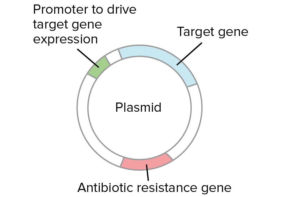
Diagram of a plasmid. The plasmid contains an antibiotic resistance gene, a promoter to drive gene expression in bacteria, and the target gene inserted during the ligation.
  1. Bacteria without a plasmid die. Each bacterium with a plasmid gives rise to a cluster of identical, plasmid-containing bacteria called a colony.
  2. Several colonies are checked to identify one with the right plasmid (e.g., by PCR or restriction digest).
  1. A colony containing the right plasmid is grown in bulk and used for plasmid or protein production.

Why do we need to check colonies?

The bacteria that make colonies should all contain a plasmid (which provides antibiotic resistance). However, it’s not necessarily the case that all of the plasmid-containing colonies will have the same plasmid.
How does that work? When we cut and paste DNA, it's often possible for side products to form, in addition to the plasmid we intend to build. For instance, when we try to insert a gene into a plasmid using a particular restriction enzyme, we may get some cases where the plasmid closes back up (without taking in the gene), and other cases where the gene goes in backwards.


Left: gene goes into plasmid forwards (pointing in the same direction as the promoter sequence). This is the desired plasmid from the ligation.
Middle: plasmid closes back up without taking in the gene. This is not a useful plasmid.
Right: gene goes into plasmid backwards (pointing back towards the promoter sequence). This is not a useful plasmid if we want to express the gene in bacteria.
Let's say we are trying to insert a gene into a plasmid so it can be expressed in bacteria. In order to do so, we must "paste" the gene into the plasmid next to the promoter, pointing in the forward direction:

Starting materials:
  • Target gene digested at both ends with a particular restriction enzyme.
  • Plasmid cut with the same restriction enzyme at a site following a promoter for bacterial expression. The promoter "points" towards the right, meaning that it will drive transcription of the DNA sequence that lies to the right.
What we want to get is:
  • A recombinant plasmid where the target gene is inserted after the promoter, pointing in the forward direction (oriented so that it's transcribed to make an mRNA that specified the desired protein).
Suppose we cut our gene and plasmid with the same enzyme and join the fragments together with DNA ligase. In some cases, the plasmid DNA and the gene DNA will combine in the right way and form the plasmid we're looking for. In other cases, though, the plasmid may simply close back up (without taking in the gene), or the gene may go into the plasmid backwards. A backwards gene cannot be expressed in bacteria to make a protein.

Left: gene goes into plasmid forwards (pointing in the same direction as the promoter sequence). This is the desired plasmid from the ligation.
Middle: plasmid closes back up without taking in the gene. This is not a useful plasmid.
Right: gene goes into plasmid backwards (pointing back towards the promoter sequence). This is not a useful plasmid if we want to express the gene in bacteria.
A ligation involves many fragments of DNA (billions of copies of the plasmid, and billions of copies of the gene). Thus, in every ligation, we will get some number of "good" plasmids and some number of "bad" ones. Each colony starts from a single bacterium with a single plasmid, so all the bacteria in a colony with have the same plasmid (either "good" or "bad").
See the article on restriction enzymes and DNA ligase for a more concrete example of how and why these different ligation products can form.
Why does it matter if a gene goes into a plasmid backwards? In some cases, it doesn't. However, if we want to express the gene in bacteria to make a protein, the gene must point in the right direction relative to the promoter, or control sequence that drives gene expression. If the gene were backwards, the wrong strand of DNA would be transcribed and no protein would be made.
Because of these possibilities, it's important to collect plasmid DNA from each colony and check to see if it matches the plasmid we were trying to build. Restriction digests, PCR, and DNA sequencing are commonly used to analyze plasmid DNA from bacterial colonies.

Protein production in bacteria

Suppose that we identify a colony with a "good" plasmid. What happens next? What's the point of all that transforming, selecting, and analyzing?

Possibility 1: Bacteria = plasmid factories

In some cases, bacteria are simply used as "plasmid factories," making lots of plasmid DNA. The plasmid DNA might be used in further DNA cloning steps (e.g., to build more complex plasmids) or in various types of experiments.
In some cases, plasmids are directly used for practical purposes. For instance, plasmids were used to deliver a human gene to lung tissue in a recent gene therapy clinical trial for patients with the genetic disorder cystic fibrosisstart superscript, 1, end superscript.

Possibility 2: Bacteria = protein factories

In other cases, bacteria may be used as protein factories. If a plasmid contains the right control sequences, bacteria can be induced to express the gene it contains when a chemical signal is added. Expression of the gene leads to production of mRNA, which is translated into protein. The bacteria can then be lysed (split open) to release the protein.


A chosen colony is grown up into a large culture. The bacteria in the large culture are induced to express the target gene through addition of a chemical signal to the culture medium. Inside each bacterium, the target gene is transcribed into mRNA, and the mRNA is translated into protein. The protein encoded by the target gene accumulates inside the bacteria.
Bacteria contain many proteins and macromolecules. Because of this, the newly made protein needs to be purified (separated from the other proteins and macromolecules) before it can be used. There are a variety of different techniques used for protein purification.

Cells that have produced protein are burst open (lysed), releasing the protein and the other cell contents. The molecules extracted from the cells are applied to a column that contains antibodies specific for the target protein. Thus, the protein is trapped in the column while other molecules from the bacteria flow through. In the final step, after all the non-target proteins have been washed away, the target proteins are released from the antibodies in the column, and the pure protein is collected for use.
In one technique called affinity chromatography, a mixture of molecules extracted from the lysed bacteria is poured through a column, or a cylinder packed with beads. The beads are coated with an antibody, an immune system protein that binds specifically to a target molecule.

The antibody in the column is designed to bind to our protein of interest, and not to any other molecules in the mixture. Thus, the protein of interest is trapped in the column, while the other molecules are washed away. In the final step, the protein of interest is released from the column and collected for use.

[Source: Khan Academy]