Tuesday, September 26, 2017

If the RNA in our bodies is essentially copied from our DNA, why might we want to study RNA instead?

A neuron in your brain and a macrophage in your blood both have the same DNA, yet their shapes, sizes, and functions are vastly different from each other.
The cells in our bodies become structurally and functionally diverse by activating different combinations of genes. By studying the RNA that is transcribed from these genes, we can find out which genes are active in a particular cell type, bringing us closer to understanding how a cell can perform its specialized job. In addition to comparing the expressed (ie. active) genes between different types of cells, we can also study how these patterns of gene expression change over time or in response to different stimuli. Using this information, we can start answering questions like "Why does taking aspirin relieve pain, and how does it cause its side effects?"
In short, examining DNA provides us with a static picture of what a cell or organism might do or become, whereas measuring RNA lets us see what a cell/organism is actually doing right now. None of this is to say sequencing RNA is "better" or more important than sequencing DNA. The truth is these two processes are dependent upon and inform each other.

Sunday, May 7, 2017

DNA, genes and chromosomes

Your genes are part of what makes you the person you are. You are different from everyone alive now and everyone who has ever lived.

DNA

Your genes also mean that you probably look a bit like other members of your family. For example, have you been told that you have 'your mother's eyes' or 'your grandmother's nose'?

Genes influence what we look like on the outside and how we work on the inside. They contain the information our bodies need to make chemicals called proteins. Proteins form the structure of our bodies, as well playing an important role in the processes that keep us alive.

Genes are made of a chemical called DNA, which is short for 'deoxyribonucleic acid'. The DNA molecule is a double helix: that is, two long, thin strands twisted around each other like a spiral staircase.
The sides are sugar and phosphate molecules. The rungs are pairs of chemicals called 'nitrogenous bases', or 'bases' for short.

There are four types of base: adenine (A), thymine (T), guanine (G) and cytosine (C). These bases link in a very specific way: A always pairs with T, and C always pairs with G.

The DNA molecule has two important properties.
  • It can make copies of itself. If you pull the two strands apart, each can be used to make the other one (and a new DNA molecule).
  • It can carry information. The order of the bases along a strand is a code - a code for making proteins.

Genes

A gene is a length of DNA that codes for a specific protein. So, for example, one gene will code for the protein insulin, which is important role in helping your body to control the amount of sugar in your blood.

Genes are the basic unit of genetics. Human beings have 20,000 to 25,000 genes. These genes account for only about 3 per cent of our DNA. The function of the remaining 97 per cent is still not clear, although scientists think it may have something to do with controlling the genes.

Chromosomes

If you took the DNA from all the cells in your body and lined it up, end to end, it would form a strand 6000 million miles long (but very, very thin)! To store this important material, DNA molecules are tightly packed around proteins called histones to make structures called chromosomes.
Human beings have 23 pairs of chromosomes in every cell, which makes 46 chromosomes in total. A photograph of a person's chromosomes, arranged according to size, is called a karyotype.
The sex chromosomes determine whether you are a boy (XY) or a girl (XX). The other chromosomes are called autosomes.
The karyotype of a male human being
The largest chromosome, chromosome 1, contains about 8000 genes. The smallest chromosome, chromosome 21, contains about 300 genes. (Chromosome 22 should be the smallest, but the scientists made a mistake when they first numbered them!).

The DNA that contains your genes is stored in your cells in a structure called the nucleus.

A diagram of animal cell showing the nucleus
Source: University of Leicester

Saturday, April 15, 2017

Microscopic effect after stroke

We talk a lot about strokes in a clinical way in medical school. We discuss which areas of the brain are involved, and we correlate the areas damaged with the patient’s symptoms.
But what actually happens in the affected brain regions after a stroke? Injuries in the brain don’t heal like they do in other organs (you don’t form a scab and a scar in your brain). Let’s take a look at the steps the body takes to heal itself following an ischemic event in the brain.
There are basically four stages of healing following an infarct, and they usually happen in a predictable timeframe.
1. The first day (12-24 hours)
  • After brain tissue dies, it takes a while before you can see any real changes in the cells. The first changes occur in neurons. Somewhere around 12 hours following an infarct, neuronal cytoplasm develops tiny holes (microvacuoles) and takes on a deep pink-red color (the neurons are actually called red neurons at this point – you can see why in the image above). 
  • Later the nucleus undergoes pyknosis (in which it becomes small and dark) and karyorrhexis (in which it fragments into little bits, like cookie crumbs. Pathologists love food analogies and use them whenever possible.) Cells in general (but especially endothelial cells and astrocytes) tend to swell up and become more faded in color. Myelinated fibers disintegrate.

2. The second day (24-48 hours)
  • Somewhere around the end of the first day, neutrophils swarm into the area, staying until about the end of the second day, at which point they take off and are replaced by  macrophages (which come in from the blood as monocytes). Microglia (the resident phagocytic cells of the brain) become activated too. 
  • The tissue begins to undergo liquefactive necrosis from all those nasty enzymes released by the neutrophils. 
  • Macrophages are like little moms going around and cleaning up the seemingly never-ending mess. 
  • Astrocytes start to react, becoming large and getting ready to divide.

3. The next few weeks (2-3 weeks) 
  • Macrophages continue to clean stuff up. They become stuffed with debris, and you can still see some of them hanging around months or even years later. 
  • Astrocytes multiply and develop prominent, arborizing cytoplasmic extensions.

4. After several months
  • Eventually, the astrocytes calm down, and what’s left is a cavity surrounded by a dense network of glial fibers and new blood vessels. There’s no collagen formation like there is in many other organs (like skin) – so there’s no filling in of the lost tissue space.

This whole process takes place from the outside of the lesion moving inward. Which is kind of cool because you’ll often see several stages of healing going on in the same lesion.
Ref.: Pathology Student

Wednesday, February 8, 2017

Bacterial Transformation & Selection

Transfer of plasmid DNA into bacteria. How bacteria are selected. Protein production and purification.


Key points:

  • Bacteria can take up foreign DNA in a process called transformation.
  • Transformation is a key step in DNA cloning. It occurs after restriction digest and ligation and transfers newly made plasmids to bacteria.
  • After transformation, bacteria are selected on antibiotic plates. Bacteria with a plasmid are antibiotic-resistant, and each one will form a colony.
  • Colonies with the right plasmid can be grown to make large cultures of identical bacteria, which are used to produce plasmid or make protein.

The big picture: DNA cloning

Transformation and selection of bacteria are key steps in DNA cloning. DNA cloning is the process of making many copies of a specific piece of DNA, such as a gene. The copies are often made in bacteria.
In a typical cloning experiment, researchers first insert a piece of DNA, such as a gene, into a circular piece of DNA called a plasmid. This step uses restriction enzymes and DNA ligase and is called a ligation.
After a ligation, the next step is to transfer the DNA into bacteria in a process called transformation. Then, we can use antibiotic selection and DNA analysis methods to identify bacteria that contain the plasmid we’re looking for.

Steps of bacterial transformation and selection

Here is a typical procedure for transforming and selecting bacteria:


  1. Specially prepared bacteria are mixed with DNA (e.g., from a ligation).
  2. The bacteria are given a heat shock, which "encourages" them to take up a plasmid. Most bacteria do not take up a plasmid, but some do.
  3. Plasmids used in cloning contain an antibiotic resistance gene. Thus, all of the bacteria are placed on an antibiotic plate to select for ones that took up a plasmid.
  4. Bacteria without a plasmid die. Each bacterium with a plasmid gives rise to a cluster of identical, plasmid-containing bacteria called a colony. A typical colony looks like a small, whitish dot the size of a pinhead.
  5. Several colonies are checked to identify one with the right plasmid.
  6. A colony containing the right plasmid is grown in bulk and used for plasmid or protein production.
  1. Specially prepared bacteria are mixed with DNA (e.g., from a ligation).
  2. The bacteria are given a heat shock, which causes some of them to take up a plasmid.
    The basic answer is that a heat shock makes the bacterial membrane more permeable to DNA molecules, such as plasmids. It appears that the heat shock causes the formation of pores in the bacterial membrane, through which the DNA molecules can pass.
  3. Plasmids used in cloning contain an antibiotic resistance gene. Thus, all of the bacteria are placed on an antibiotic plate to select for ones that took up a plasmid.

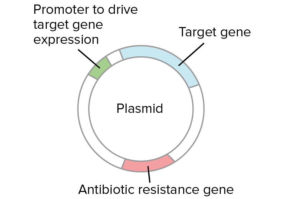
Diagram of a plasmid. The plasmid contains an antibiotic resistance gene, a promoter to drive gene expression in bacteria, and the target gene inserted during the ligation.
  1. Bacteria without a plasmid die. Each bacterium with a plasmid gives rise to a cluster of identical, plasmid-containing bacteria called a colony.
  2. Several colonies are checked to identify one with the right plasmid (e.g., by PCR or restriction digest).
  1. A colony containing the right plasmid is grown in bulk and used for plasmid or protein production.

Why do we need to check colonies?

The bacteria that make colonies should all contain a plasmid (which provides antibiotic resistance). However, it’s not necessarily the case that all of the plasmid-containing colonies will have the same plasmid.
How does that work? When we cut and paste DNA, it's often possible for side products to form, in addition to the plasmid we intend to build. For instance, when we try to insert a gene into a plasmid using a particular restriction enzyme, we may get some cases where the plasmid closes back up (without taking in the gene), and other cases where the gene goes in backwards.


Left: gene goes into plasmid forwards (pointing in the same direction as the promoter sequence). This is the desired plasmid from the ligation.
Middle: plasmid closes back up without taking in the gene. This is not a useful plasmid.
Right: gene goes into plasmid backwards (pointing back towards the promoter sequence). This is not a useful plasmid if we want to express the gene in bacteria.
Let's say we are trying to insert a gene into a plasmid so it can be expressed in bacteria. In order to do so, we must "paste" the gene into the plasmid next to the promoter, pointing in the forward direction:

Starting materials:
  • Target gene digested at both ends with a particular restriction enzyme.
  • Plasmid cut with the same restriction enzyme at a site following a promoter for bacterial expression. The promoter "points" towards the right, meaning that it will drive transcription of the DNA sequence that lies to the right.
What we want to get is:
  • A recombinant plasmid where the target gene is inserted after the promoter, pointing in the forward direction (oriented so that it's transcribed to make an mRNA that specified the desired protein).
Suppose we cut our gene and plasmid with the same enzyme and join the fragments together with DNA ligase. In some cases, the plasmid DNA and the gene DNA will combine in the right way and form the plasmid we're looking for. In other cases, though, the plasmid may simply close back up (without taking in the gene), or the gene may go into the plasmid backwards. A backwards gene cannot be expressed in bacteria to make a protein.

Left: gene goes into plasmid forwards (pointing in the same direction as the promoter sequence). This is the desired plasmid from the ligation.
Middle: plasmid closes back up without taking in the gene. This is not a useful plasmid.
Right: gene goes into plasmid backwards (pointing back towards the promoter sequence). This is not a useful plasmid if we want to express the gene in bacteria.
A ligation involves many fragments of DNA (billions of copies of the plasmid, and billions of copies of the gene). Thus, in every ligation, we will get some number of "good" plasmids and some number of "bad" ones. Each colony starts from a single bacterium with a single plasmid, so all the bacteria in a colony with have the same plasmid (either "good" or "bad").
See the article on restriction enzymes and DNA ligase for a more concrete example of how and why these different ligation products can form.
Why does it matter if a gene goes into a plasmid backwards? In some cases, it doesn't. However, if we want to express the gene in bacteria to make a protein, the gene must point in the right direction relative to the promoter, or control sequence that drives gene expression. If the gene were backwards, the wrong strand of DNA would be transcribed and no protein would be made.
Because of these possibilities, it's important to collect plasmid DNA from each colony and check to see if it matches the plasmid we were trying to build. Restriction digests, PCR, and DNA sequencing are commonly used to analyze plasmid DNA from bacterial colonies.

Protein production in bacteria

Suppose that we identify a colony with a "good" plasmid. What happens next? What's the point of all that transforming, selecting, and analyzing?

Possibility 1: Bacteria = plasmid factories

In some cases, bacteria are simply used as "plasmid factories," making lots of plasmid DNA. The plasmid DNA might be used in further DNA cloning steps (e.g., to build more complex plasmids) or in various types of experiments.
In some cases, plasmids are directly used for practical purposes. For instance, plasmids were used to deliver a human gene to lung tissue in a recent gene therapy clinical trial for patients with the genetic disorder cystic fibrosisstart superscript, 1, end superscript.

Possibility 2: Bacteria = protein factories

In other cases, bacteria may be used as protein factories. If a plasmid contains the right control sequences, bacteria can be induced to express the gene it contains when a chemical signal is added. Expression of the gene leads to production of mRNA, which is translated into protein. The bacteria can then be lysed (split open) to release the protein.


A chosen colony is grown up into a large culture. The bacteria in the large culture are induced to express the target gene through addition of a chemical signal to the culture medium. Inside each bacterium, the target gene is transcribed into mRNA, and the mRNA is translated into protein. The protein encoded by the target gene accumulates inside the bacteria.
Bacteria contain many proteins and macromolecules. Because of this, the newly made protein needs to be purified (separated from the other proteins and macromolecules) before it can be used. There are a variety of different techniques used for protein purification.

Cells that have produced protein are burst open (lysed), releasing the protein and the other cell contents. The molecules extracted from the cells are applied to a column that contains antibodies specific for the target protein. Thus, the protein is trapped in the column while other molecules from the bacteria flow through. In the final step, after all the non-target proteins have been washed away, the target proteins are released from the antibodies in the column, and the pure protein is collected for use.
In one technique called affinity chromatography, a mixture of molecules extracted from the lysed bacteria is poured through a column, or a cylinder packed with beads. The beads are coated with an antibody, an immune system protein that binds specifically to a target molecule.

The antibody in the column is designed to bind to our protein of interest, and not to any other molecules in the mixture. Thus, the protein of interest is trapped in the column, while the other molecules are washed away. In the final step, the protein of interest is released from the column and collected for use.

[Source: Khan Academy]

Sunday, January 29, 2017

Neurons, Synapses, Action Potentials, and Neurotransmission

Function of neurons

The central nervous system [CNS] is composed entirely of two kinds of specialized cells: neurons and glia. Hence, every information processing system in the CNS is composed of neurons and glia; so too are the networks that compose the systems (and the maps). Clearly, without these two types of cells, the CNS would not be able to do what it does (which is everything having to do with our minds and how we move our bodies). But what do neurons and glia themselves do? What are their functions?
Neurons are the basic information processing structures in the CNS. Everything occurring above the level of neurons qualifies as information processing too. But nothing below the level of neurons does. We shall ignore that this view, called the neuron doctrine, is somewhat controversial. What isn't controversial is that the function of a neuron is to receive INPUT "information" from other neurons, to process that information, then to send "information" as OUTPUT to other neurons. (Synapses are connections between neurons through which "information" flows from one neuron to another.) Hence, neurons process all of the "information" that flows within, to, or out of the CNS. All of it! All of the motor information through which we are able to move; all of the sensory information through which we are able to see, to hear, to smell, to taste, and to touch; and of course all of the cognitive information through which we are able to reason, to think, to dream, to plan, to remember, and to do everything else that we do with our minds. Processing so many kinds of information requires many types of neurons; there may be as many as 10,000 types of them. Processing so much information requires a lot of neurons. How many? Well, "best estimates" indicate that there are around 200 billion neurons in the brain alone! And as each of these neurons is connected to between 5,000 and 200,000 other neurons, the number of ways that information flows among neurons in the brain is so large, it is greater than the number stars in the entire universe!
While we are considering numbers, it is worth noting that there are as many as 50 times more glia than neurons in our CNS! Glia (or glial cells) are the cells that provide support to the neurons. In much the same way that the foundation, framework, walls, and roof of a house prove the structure through which run various electric, cable, and telephone lines, along with various pipes for water and waste, not only do glia provide the structural framework that allows networks of neurons to remain connected, they also attend to the brain's various house keeping functions (such as removing debris after neuronal death).
Because our main interest lies in exploring how information processing occurs in the brain, we are going to ignore glia. But before we see how neurons process information (and what that means), you need to know a few things about the structure of neurons.

Structure of neurons

While there are as many as 10,000 specific types of neurons in the human brain, generally speaking, there are three kinds of neurons: motor neurons (for conveying motor information), sensory neurons (for conveying sensory information), and interneurons (which convey information between different types of neurons). The following image identifies how neurons come in various shapes and sizes. (It is based on drawings made by Cajal.)
A "typical" neuron has four distinct parts (or regions). The first part is the cell body (or soma). This is not only the metabolic "control center" of the neuron, it is also its "manufacturing and recycling plant." (For instance, it is within the cell body that neuronal proteins are synthesized.) The second and third parts are processes — structures that extend away from the cell body. Generally speaking, the function of a process is to be a conduit through which signals flow to or away from the cell body. Incoming signals from other neurons are (typically) received through its dendrites. The outgoing signal to other neurons flows along its axon. A neuron may have many thousands of dendrites, but it will have only one axon. The fourth distinct part of a neuron lies at the end of the axon, the axon terminals. These are the structures that contain neurotransmitters. Neurotransmitters are the chemical medium through which signals flow from one neuron to the next at chemical synapses.

Neuronal signaling

To support the general function of the nervous system, neurons have evolved unique capabilities for intracellular signaling (communication within the cell) and intercellular signaling (communication between cells). To achieve long distance, rapid communication, neurons have evolved special abilities for sending electrical signals (action potentials) along axons. This mechanism, called conduction, is how the cell body of a neuron communicates with its own terminals via the axon. Communication between neurons is achieved at synapses by the process of neurotransmission.

Conduction

To begin conduction, an action potential is generated near the cell body portion of the axon. An action potential is an electrical signal very much like the electrical signals in electronic devices. But whereas an electrical signal in an electronic device occurs because electrons move along a wire, an electrical signal in a neuron occurs because ions move across the neuronal membrane. Ions are electrically charged particles. The protein membrane of a neuron acts as a barrier to ions. Ions move across the membrane through ion channels that open and close due to the presence of neurotransmitter. When the concentration of ions on the inside of the neuron changes, the electrical property of the membrane itself changes. Normally, the membrane potential of a neuron rests as -70 millivolts (and the membrane is said to be polarized). The influx and outflux of ions (through ion channels during neurotransmission) will make the inside of the target neuron more positive (hence, de-polarized). When this depolarization reaches a point of no return called a threshold, a large electrical signal is generated. This is the action potential. How it is generated is illustrated in the following animation.
This signal is then propagated along the axon (and not, say, back to its dendrites) until it reaches its axon terminals. An action potential travels along the axon quickly, moving at rates up to 150 meters (or roughly 500 feet) per second. Conduction ends at the axon terminals. Axon terminals are where neurotransmission begins. Hence, it is at axon terminals where the neuron sends its OUTPUT to other neurons. At electrical synapses, the OUTPUT will be the electrical signal itself. At chemical synapses, the OUTPUT will be neurotransmitter.

Neurotransmission

Neurotransmission (or synaptic transmission) is communication between neurons as accomplished by the movement of chemicals or electrical signals across a synapse. For any interneuron, its function is to receive INPUT "information" from other neurons through synapses, to process that information, then to send "information" as OUTPUT to other neurons through synapses. Consequently, an interneuron cannot fulfill its function if it is not connected to other neurons in a network. A network of neurons (or neural network) is merely a group of neurons through which information flows from one neuron to another. The image below represents a neural network. "Information" flows between the blue neurons through electrical synapses. "Information" flows from yellow neuron A, through blue neuron B, to pink neuron C via chemical synapses.


The following animation illustrates the difference between these two kinds of synapses.

At electrical synapses, two neurons are physically connected to one another through gap junctions. Gap junctions permit changes in the electrical properties of one neuron to effect the other, and vice versa, so the two neurons essentially behave as one. Electrical neurotransmission is communication between two neurons at electrical synapses. How this occurs is explored in a bit more detail in the following animation.
Chemical neurotransmission occurs at chemical synapses. In chemical neurotransmission, the presynaptic neuron and the postsynaptic neuron are separated by a small gap — the synaptic cleft. The synaptic cleft is filled with extracellular fluid (the fluid bathing all the cells in the brain). Although very small, typically on the order of a few nanometers (a billionth of a meter), the synaptic cleft creates a physical barrier for the electrical signal carried by one neuron to be transferred to another neuron. In electrical terms, the synaptic cleft would be considered a “short” in an electrical circuit. The function of neurotransmitter is to overcome this electrical short. It does so by acting like a chemical messenger, thereby linking the action potential of one neuron with a synaptic potential in another. How this occurs is illustrated in the following animation.