Neurulation
 Neural Tube
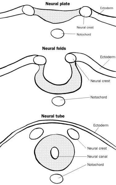
The nervous system develops when the notochord induces its overlying ectoderm to become neuroectoderm and to develop into the neural plate. The neural plate folds along its central axis to form a neural groove lined on each side by a neural fold. The two neural folds fuse together and pinch off to become the neural tube. Fusion of the neural folds begins in the middle of the embryo and moves cranially and caudally. The cranial open end of the tube is the anterior (rostral) neuropore, and the caudal open end of the tube is the posterior (caudal) neuropore. The anterior neuropore closes on or before day 26 and the caudal neuropore closes before the end of the fourth week.
Neural Crest
Some cells from the neural folds give rise to pleuripotent neural crest cells that migrate widely in the embryo and give rise to many nervous structures:
· Spinal ganglia (dorsal root ganglia)
· Ganglia of the autonomic nervous system
· Ganglia of some cranial nerves
· Sheaths of peripheral nerves
· Meninges of brain and spinal cord
· Pigment cells
· Suprarenal medulla
· Skeletal and muscular components in the head
Central Nervous System
Development of Brain
The neural tube forms three primary brain vesicles. The primary brain vesicles give rise to five secondary brain vesicles, which give rise to various adult structures.
Primary vesicles
|
Secondary vesicles
|
Adult structures
|
|
Forebrain vesicle (prosencephalon)
|
Telencephalon
|
Cerebral hemispheres, consisting of the cortex and medullary center, basal ganglia, lamina terminalis, hippocampus, the corpus striatum, and the olfactory system
|
| |
Diencephalon
|
Thalamus, epithalamus, hypothalamus, subthalamus, neurohypophysis, pineal gland, retina, optic nerve, mamillary bodies
|
|
Midbrain vesicle (mesencephalon)
|
Mesencephalon
|
Midbrain
|
|
Hindbrain vesicle (rhombencephalon)
|
Metencephalon
|
Pons and cerebellum
|
| |
Myelencephalon
|
Medulla
|
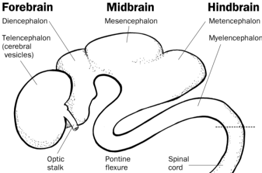
Figure - Structure of embryonic brain
Development of Spinal Cord
The neural tube consists of three cellular layers from inner to outer: the ventricular zone (ependymal layer), the intermediate zone (mantle layer), and the marginal zone (marginal layer). The ventricular zone gives rise to neuroblasts (future nerve cells) and glioblasts (future supporting cells) which migrate into the intermediate zone form two collections of cells (the alar plate and the basal plate) separated by a groove called the sulcus limitans. Cells in the alar plate become afferent (sensory) neurons and form the dorsal (posterior) horn of the spinal cord. Cells in the basal plate become efferent (motor) neurons and form the ventral (anterior) horn of the spinal cord. The two ventral horns bulge ventrally to create ventral median fissure. The dorsal horns merge to create the dorsal median septum. The lumen of the neural tube becomes the central canal of the spinal cord.
The spinal cord extends the entire length of the vertebral canal at week 8 of development. At birth, the conus medullaris extends to the L3 vertebra. In the adult, the conus medullaris extends to the L1 vertebra. Spinal lumbar punctures must be performed caudally to the conus medullaris to avoid damaging the spinal cord.
Development of Meninges
The dura mater arises from paraxial mesoderm that surrounds the neural tube. The pia mater and arachnoid mater arise from neural crest cells.
Pituitary Gland
The anterior pituitary gland (adenohypophysis) arises from an evagination of the oropharyngeal membrane known as Rathke’s pouch. The posterior pituitary gland (neurohypophysis) arises from an evagination of neuroectoderm from the diencephalon.
Clinical Correlations
Spina Bifida
· Spina bifida occulta is a defect of the vertebral column only, and is a common problem affecting as many as 10% of live births.
· Spina bifida with meningocele (spina bifida cystica) is a defect of the vertebral column with protrusion of the meninges through the defect.
· Spina bifida with myelomeningocele is a defect of the vertebral column protrusion of the meninges and herniation of the spinal cord through the defect.
· Spina bifida with myeloschisis results from the failure of the caudal neuropore to close at the end of the fourth week of development. Newborn infants are paralyzed distal to the lesion.
These defects usually occur in the cervical and/or lumbar regions and may cause neurologic deficits in the lower limbs and urinary bladder. Neural tube defects can be detected by the presence of alpha-fetoprotein (AFP) in the fetal circulation after the fourth week of development.
Anencephaly
Anencephaly is the failure of the anterior neuropore to close, resulting in a failure of the brain to develop.
Microcephaly
Microcephaly (small head) results from microencephaly (small brain), or the failure of the brain to grow normally. This can be the result of exposure to large doses of radiation up to the sixteenth week of development, or from certain infectious agents (cytomegalovirus, herpes simplex virus, and toxoplasma gondii)
Hydrocephalus
Hydrocephalus is an accumulation of CSF in the ventricles of the brain, caused most commonly by stenosis of the cerebral aqueduct. In the absence of surgical treatment in extreme cases the head may swell to three times its normal size.
Arnold-Chiari Malformation
Arnold-Chiari malformation is herniation parts of the cerebellum (medulla oblongata and cerebellar vermis) through the foramen magnum of the skull.
Fetal Alcohol Syndrome
Ingestion of alcohol during pregnancy is the most common cause of infant mental retardation. It also causes microcephaly and congenital heart disease.
[Source: University of Michigan Medical School]The Cerebrum: The cerebrum or cortex is the largest part of the human brain, associated with higher brain function such as thought and action. The cerebral cortex is divided into four sections, called "lobes": frontal lobe, parietal lobe, occipital lobe, and temporal lobe. Here is a visual representation of the cortex:

- Frontal Lobe- associated with reasoning, planning, parts of speech, movement, emotions, and problem solving
- Parietal Lobe- associated with movement, orientation, recognition, perception of stimuli
- Occipital Lobe- associated with visual processing
- Temporal Lobe- associated with perception and recognition of auditory stimuli, memory, and speech
A deep furrow divides the cerebrum into two halves, known as the left and right hemispheres. The two hemispheres look mostly symmetrical yet it has been shown that each side functions slightly different than the other. Sometimes the right hemisphere is associated with creativity and the left hemispheres is associated with logic abilities. The corpus callosum is a bundle of axons which connects these two hemispheres.
Nerve cells make up the gray surface of the cerebrum which is a little thicker than your thumb. White nerve fibers underneath carry signals between the nerve cells and other parts of the brain and body.
The neocortex occupies the bulk of the cerebrum. This is a six-layered structure of the cerebral cortex which is only found in mammals. It is thought that the neocortex is a recently evolved structure, and is associated with "higher" information processing by more fully evolved animals (such as humans, primates, dolphins, etc).
The Cerebellum: The cerebellum, or "little brain", is similar to the cerebrum in that it has two hemispheres and has a highly folded surface or cortex. This structure is associated with regulation and coordination of movement, posture, and balance.
Limbic System: The limbic system, often referred to as the "emotional brain", is found buried within the cerebrum. Like the cerebellum, evolutionarily the structure is rather old.
This system contains thalamus, hypothalamus, amygdala, and hippocampus. Here is a visual representation of this system, from a midsagittal view of the human brain:

Thalamus- a large mass of gray matter deeply situated in the forebrain at the topmost portion of the diencephalon. The structure has sensory and motor functions. Almost all sensory information enters this structure where neurons send that information to the overlying cortex. Axons from every sensory system (except olfaction) synapse here as the last relay site before the information reaches the cerebral cortex.

Hypothalamus- part of the diencephalon, ventral to the thalamus. The structure is involved in functions including homeostasis, emotion, thirst, hunger, circadian rhythms, and control of the autonomic nervous system. In addition, it controls the pituitary.

Amygdala- part of the telencephalon, located in the temporal lobe; involved in memory, emotion, and fear. The amygdala is both large and just beneath the surface of the front, medial part of the temporal lobe where it causes the bulge on the surface called the uncus. This is a component of the limbic system.

Hippocampus- the portion of the cerebral hemispheres in basal medial part of the temporal lobe. This part of the brain is important for learning and memory . . . for converting short term memory to more permanent memory, and for recalling spatial relationships in the world about us

The brain stem is made of midbrain, pons, and medulla.
Midbrain/ Mesencephalon- the rostral part of the brain stem, which includes the tectum and tegmentum. It is involved in functions such as vision, hearing, eye movement, and body movement. The anterior part has the cerebral peduncle, which is a huge bundle of axons traveling from the cerebral cortex through the brain stem and these fibers (along with other structures) are important for voluntary motor function.

Pons- part of the metencephalon in the hindbrain. It is involved in motor control and sensory analysis... for example, information from the ear first enters the brain in the pons. It has parts that are important for the level of consciousness and for sleep. Some structures within the pons are linked to the cerebellum, thus are involved in movement and posture.

Medulla Oblongata- this structure is the caudal-most part of the brain stem, between the pons and spinal cord. It is responsible for maintaining vital body functions, such as breathing and heartrate

No comments:
Post a Comment